NSJ Bioreagents
![]()
NSJ Bioreagents(後稱NSJBio)在抗體製作領域上已經有超過20年以上的豐厚經驗。NSJBio除了提供傳統常用的各種單株抗體外,也同時提供新入市場的嶄新前沿單株/多株抗體,重組單株抗體,以及通過人體蛋白微陣列驗證的抗體。產品涵蓋癌症研究,腫瘤生物標記,細胞凋亡,免疫學,細胞生物,轉譯因子,表觀遺傳學等多項領域。為了對應不同實驗室的使用需求及經費預算,多數產品都有多種大小可供選擇。NSJBio相信在他們嚴謹的品質把關下,絕對能提供給您高品質且可信賴的產品。
以下為NSJBio全球熱銷產品的介紹:

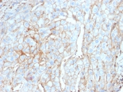
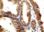
PD-L1 Antibody / B7-H1 / CD274 [clone PDL1/2746] (V3955)
當抗原存在時,CD28和B7-1(CD80)或B7-2(CD86)的結合會刺激T細胞的增生,細胞因子的產生,作用性T細胞的分化,以及BCLX(一種T細胞存活因子)的誘發。而B7-1或B7-2所誘引來的CTLA4則有可能限制細胞增生及白細胞介素2(IL-2)的生成。PD-L1是一個290氨基酸的第一型跨膜蛋白,結構上和B7-1有20%相似,和B7-1有15%相似。PD-L1具有類V及類C抗體結構域和30氨基酸長的細胞質領域。PD-L1不會和CD28,細胞毒性T細胞A4,或可誘導共激活分子結合。IL-2雖然生產量不高,卻是PD-L1共激活效果所必要的分子。PD-L2蛋白含有一個信號序列,類V及類C抗體領域,跨膜領域,以及細胞質領域。在心臟,肺,以及腎臟的薄壁組織常態性表示PD-L1和PD-L2的實驗結果可能指出PD-1-PD-L系統可能可以透過發出負面信號藉以幫助預防自體免疫疾病。
更多資料請參考本項產品的產品資料表

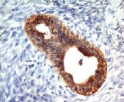
Histone Antibody / Histone H1 [clone 1415-1] (V2567)
真核動物的組蛋白是鹼性的水溶性核蛋白,通過左旋包覆146對核酸來形成染色體纖維並組成異八聚體核小體顆粒。八聚體為四個核心組蛋白(H2A, H2B, H3, 和H4)各兩個組成(兩個H2A-H2B二聚體及兩個H3-H4二聚體組成一個幾乎完全對稱的三級結構)。超過80%的核小體含有組蛋白H1,其來源自不含内含子的基因。該基因會與核小體之間的基因互動,並引導基因壓縮成染色質。組蛋白會受酶對其N末端的修改以及對其球狀結構的修改的影響。這類修改包括添加甲基,瓜氨酸,乙酰,磷酸,SUMO蛋白,泛素,和ADP-核糖基。
更多資料請參考本項產品的產品資料表

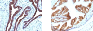
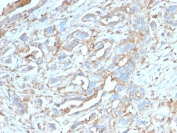
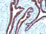
E-Cadherin Antibody [clone CDH1/1525] (V3190)
這個抗體針對的是80-120kDa的E-鈣粘蛋白。鈣粘蛋白是由一系列依賴鈣離子的黏附分子組成,其功能是引導對維持組織結構和形態發生至關重要的細胞-細胞閒的粘合。傳統的鈣粘蛋白E-, N-, 和P-鈣粘蛋白是由具有5個同源的NH2末端重複的細胞外結構域組成。其較短的細胞内結構域與多種細胞質蛋白(如β-連環蛋白)相互作用來調節鈣粘蛋白的功能。E-鈣粘蛋白在上皮細胞的黏附上也起到了重要的作用。E-鈣粘蛋白的降低表現被推斷和乳腺癌,前列腺癌,和食道癌的轉移潛能和預後不良可能有關。和p120連環蛋白共同使用,可以用於分化管線(E-鈣粘蛋白+)和小葉(E-鈣粘蛋白-)乳腺癌之間的差別。其也被認為可能有助於診斷間皮瘤。
更多資料請參考本項產品的產品資料表
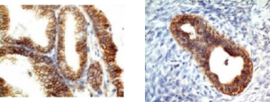
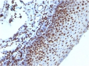
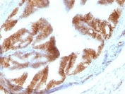
EpCAM Antibody / Cytoplasmic domain [clone EGP40/1110] (V2686)
EGP40是一個40-43kDa的跨膜上皮糖蛋白,也被認定為上皮特異性抗原(Epithelial Specific Antigen, ESA)或是上皮細胞黏附分子(Epithelial Cellular Adhesion Molecule, Ep-CAM)。其在大部分的單純上皮細胞或大部分的癌症的情況下都在基底外側細胞表面上表達。 本抗體已被用於區分腺癌,胸膜間皮癌,和肝細胞癌。 本抗體也可用於區分卵巢的漿液性癌和間皮癌。
更多資料請參考本項產品的產品資料表
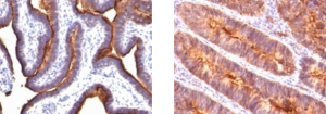
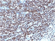
EMA Antibody / MUC1 / Mucin-1 [clone MUC1/845] (V2371)
這個抗體可以辨識在265-400kDa範圍的蛋白質,鑒定為MUC1(粘蛋白1)或EMA(上皮膜抗原)的不同糖類別。其α單位具有細胞黏附的特性。其可以同時黏附蛋白和抗黏附蛋白的作用。MUC1 / 粘蛋白1可以在上皮細胞上提供抗菌和酶攻擊的保護層。 Β單位具有C末端結構域,其可以通過磷酸化和蛋白質-蛋白質相互作用來參與細胞的信號傳導。 在免疫組織化學分析中,MUC1 / EMA抗體可以對常規的福爾馬林/石蠟癌組織進行極有效的染色。 MUC1抗體同時也可用作泛上皮標記物,用於檢測骨髓或肝臟中癌的早期轉移基因座。
更多資料請參考本項產品的產品資料表
NSJ Bioreagents

Many products are offered at multiple sizes to better fit researchers testing needs and budget constraints. Comprehensive product data sheets provide product information and testing data images and are backed up by experienced technical service. And all products are 100% guaranteed to work as stated on the product data sheets.
Here is the best sales products which are used by many researchers all over the world.
PD-L1 Antibody / B7-H1 / CD274 [clone PDL1/2746] (V3955)
Engagement of CD28 by B7-1 (CD80) or B7-2 (CD86) in the presence of antigen promotes T-cell proliferation, cytokine production, differentiation of effector T-cells and the induction of BCLX, a promoter of T-cell survival. recruitment of CTLA4 by B7-1 or B7-2, on the other hand, may inhibit proliferation and interleukin-2 (IL-2) production.
PD-L1 is 290-amino acid type I transmembrane protein, which is 20% and 15% identical to B7-1 and B7-2, respectively, has immunoglobulin V-like and C-like domains and a 30-amino acid cytoplasmic tail. PD-L1 does not bind CD28, cytotoxic T-lymphocyte A4 or ICOS (inducible co-stimulator). IL-2, although produced in small amounts, is required for the effect of PD-L1 co-stimulation. PD-L2 protein contains a signal sequence, IgV- and IgC-like domains, a transmembrane region and a cytoplasmic region.
Constitutive expression of PD-L1 and PD-L2 on parenchymal cells of heart, lung and kidney suggests that the PD-1-PD-L system could provide unique negative signaling to help prevent autoimmune diseases.
Please check Product data sheet for the detail of this product.
Histone Antibody / Histone H1 [clone 1415-1] (V2567)
Eukaryotic histones are basic and water-soluble nuclear proteins that form hetero-octameric nucleosome particles by wrapping 146 base pairs of DNA in a left-handed super-helical turn sequentially to form chromosomal fiber. Two molecules of each of the four core histones (H2A, H2B, H3, and H4) form the octamer; formed of two H2A-H2B dimers and two H3-H4 dimers, forming two nearly symmetrical halves by tertiary structure. Over 80% of nucleosomes contain the linker Histone H1, derived from an intronless gene that interacts with linker DNA between nucleosomes and mediates compaction into higher order chromatin.
Histones are subject to posttranslational modification by enzymes primarily on their N-terminal tails, but also in their globular domains. Such modifications include methylation, citrullination, acetylation, phosphorylation, sumoylation, ubiquitination and ADP-ribosylation.
Please check Product data sheet for the detail of this product.
E-Cadherin Antibody [clone CDH1/1525] (V3190)
Recognizes a protein of 80-120kDa, identified as E-cadherin. Cadherins comprise a family of Ca2+-dependent adhesion molecules that function to mediate cell-cell binding critical to the maintenance of tissue structure and morphogenesis. The classical cadherins, E-, N- and P-cadherin, consist of large extracellular domains characterized by a series of five homologous NH2 terminal repeats. The relatively short intracellular domains interact with a variety of cytoplasmic proteins, such as β-catenin, to regulate cadherin function.
E-cadherin plays an important role in epithelial cell adhesion. A decreased expression of E-cadherin is associated with metastatic potential and poor prognosis in breast cancer, prostate and esophageal cancer. In combination with p120 Catenin, it is useful for the differentiation between ductal (E-cadherin +) and lobular (E-cadherin -) breast carcinomas. It may also help in diagnosis of mesothelioma.
Please check Product data sheet for the detail of this product.
EpCAM Antibody / Cytoplasmic domain [clone EGP40/1110] (V2686)
EGP40 is a 40-43kDa transmembrane epithelial glycoprotein, also identified as epithelial specific antigen (ESA), or epithelial cellular adhesion molecule (Ep-CAM). It is expressed on baso-lateral cell surface in most simple epithelia and a vast majority of carcinomas.
This antibody has been used to distinguish adenocarcinoma from pleural mesothelioma and hepatocellular carcinoma. This antibody is also useful in distinguishing serous carcinomas of the ovary from mesothelioma.
Please check Product data sheet for the detail of this product.
EMA Antibody / MUC1 / Mucin-1 [clone MUC1/845] (V2371)
This antibody recognizes proteins in MW range of 265-400 kDa, identified as different glycoforms of MUC1 (Mucin-1) or EMA (epithelial membrane antigen). The alpha subunit has cell adhesive properties. It can act both as an adhesion and an anti-adhesion protein.
MUC1 / Mucin-1 may provide a protective layer on epithelial cells against bacterial and enzyme attack. The beta subunit contains a C-terminal domain, which is involved in cell signaling through phosphorylations and protein-protein interactions. In immunohistochemical assays, the MUC1 / EMA antibody superbly stains routine formalin/paraffin carcinoma tissues. MUC1 antibody is useful as a pan-epithelial marker for detecting early metastatic loci of carcinoma in bone marrow or liver.
Please check Product data sheet for the detail of this product.
List of antibody categories
- ABO
- Angiogenesis
- Apoptosis
- Apoptosis Marker
- Astrocyte
- Autophagy
- B-Cell
- Beta Cell
- Calcium Channel
- Cancer
- Cardiovascular
- Cell Biology
- Cell Cycle
- Cellular Marker & Epitope Tag
- Cytokine
- Dendritic Cell
- Developmental Biology
- Endoplasmic Reticulum
- Endosome
- Endosome Marker
- Endothelial
- Endothelial Cell
- Endothelial Cell Marker
- Epigenetics
- ER
- Erythrocyte
- Follicular Lymphoma
- Granuloctye
- Hairy Cell Leukemia
- Hepatocellular Carcinoma
- Hepatocyte
- Hepatocyte Marker
- Human Protein Microarray Validated
- Ig
- Immunology
- Innate Immunity
- Isotype Control
- Late Endosome Marker
- Leukocyte
- Loading Control
- Lymphocyte
- Macrophage
- Mantle Cell
- Melanoma
- Mesenchymal Cell
- Metabolism
- Microbiology
- Microglia
- Mitochondria
- Mitochondrial
- Monocyte
- Myeloid Cell
- Neuronal
- Neuronal Marker
- Neuroscience
- Neutrophil
- NF-kB
- NK Cell
- Nuclear
- Phospho
- Platelet
- Popular
- Proliferation
- Rabbit Monoclonal
- Recombinant
- Recombinant Mouse
- Renal Cell
- Signal Transduction
- Skeletal Muscle
- Smooth Muscle
- Stem Cell
- Stem Cell Marker
- T-Cell
- Thyroid
- TLR & Related
- Zinc Transporter
Protocols
NSJ Bioreagents
売れ筋製品のご紹介
PD-L1抗体 / B7-H1 / CD274 [clone PDL1/2746] (V3955)
抗原存在下でCD28がリガンドのB7-1(CD80)/B7-2(CD86)と結合すると、共刺激を介してT細胞増殖分化能及びサイトカイン産生が促進されます。またこの際、T細胞のアポトーシス抑制に関わるBcl-x因子の発現も誘導されます。一方で、活性化T細胞の表面に発現するCTLA-4はCD28よりも強くB7-1/B7-2と結合し、T細胞の活性化及びIL-2産生を抑制する方向に作用します。
PD-L1はB7-1と20%、B7-2と15%類似した290残基のアミノ酸からなるⅠ型膜貫通タンパク質で、Ig可変領域と定常領域、30アミノ酸の細胞内領域を持ちます。PD-L1単体ではCD28やCTLA-4、ICOS (induced co-stimulator)と結合できず、反応のためには共刺激分子のIL-2が必要になります。PD-L2もPD-L1と同様に膜貫通タンパク質で、Ig可変領域と定常領域、30アミノ酸の細胞内領域を持つことが知られています。
心臓、肺および腎臓の実質細胞上にはPD-L1およびPD-L2が構成的に発現しています。今後PD-1/PD-L1系の機序がより解明されれば、シグナル伝達の一部を自己免疫疾患の予防に役立てることも可能になるでしょう。
製品詳細はデータシートにてご確認ください。
ヒストン抗体 / Histone H1 [clone 1415-1] (V2567)
ヒストンは真核生物のクロマチンを構成する塩基性及び水溶性の核タンパク質で、多くはヒストン八量体の状態で存在しています。ひとつのヒストン八量体は4種類のコアヒストン(H2A・H2B・H3・H4)により構成されており、H2A-H2B二量体とH3-H4二量体のペアがそれぞれ対称的に向かい合うような三次構造をとっています。ヒストン八量体に約146塩基対のDNAが左巻きに超らせん状に巻き付くことにより、クロマチン構造の最小単位であるヌクレオソームが形成されます。一方、ヒストンH1は80%以上のヌクレオソームにリンカーヒストンとして存在し、ヌクレオソーム内のDNAと結合することによりクロマチン構造を安定させる役割を持っています。
翻訳後修飾を受けやすいのは主にヒストンテールとよばれる鎖状のN末端側ですが、球状のC末端側も修飾を受けることがあります。これらの化学修飾にはメチル化、シトルリン化、アセチル化、リン酸化、ユビキチン化、SUMO化及びADPリボシル化が含まれます。
製品詳細はデータシートにてご確認ください。
E-カドヘリン抗体 [clone CDH1/1525] (V3190)
カドヘリンはカルシウム依存性に細胞接着を媒介する分子であり、細胞組織の形態維持や形態形成において重要な役割を果たします。カドヘリンスーパーファミリーにおける古典的カドヘリンのE-、N-、P-カドヘリンは、N末端側の細胞外領域に細胞接着に関わる5つの繰り返し構造(EC1~EC5)を持ちます。C末端側の細胞内領域は比較的短めで、β-カテニンなどの細胞質に存在する種々のタンパク質と相互作用することでカドヘリンの機能を調節しています。
E-カドヘリンは特に上皮細胞の接着に関係し、その発現の低下はがんの転移や予後の悪化につながります(乳がん、前立腺がん、食道がんなど)。一方で、p-120カテニンと併用することで非浸潤性乳管がん(E-カドヘリン陽性)と上皮内小葉がん(E-カドヘリン陽性)の鑑別を行うことも可能です。また、悪性中皮腫の診断にも有用です。
製品詳細はデータシートにてご確認ください。
Ep-CAM 抗体 / 細胞質ドメイン [clone EGP40/1110] (V2686)
EGP40は40-43kDa のⅠ型膜貫通型糖タンパクであり、上皮特異的抗原(epithelial specific antigen; ESA)または上皮細胞接着分子(epithelial cellular adhesion molecule; Ep-CAM)とも呼ばれます。ほとんどの正常な単層上皮の基底膜及び側細胞膜表面上に発現している一方で、大半の癌腫にも発現します。
Ep-CAM抗体は腺がんを悪性胸膜中皮腫や肝細胞癌から鑑別する際に用いられます。また、卵巣における漿液性腺癌と上皮型中皮腫の鑑別にも有用です。
製品詳細はデータシートにてご確認ください。
EMA 抗体 / MUC1 / Mucin-1 [clone MUC1/845] (V2371)
EMA抗体はムチン-1(MUC1)または上皮膜抗原(EMA)など、複数のグリコフォームを持つ分子量265-400kDaのタンパク質を認識します。EMA抗体のαサブユニットは細胞接着部分を有し、接着タンパク質または抗接着タンパク質のどちらとしても機能します。
MUC/Mucin-1は細菌や酵素などの攻撃から上皮細胞を守る保護層を形成します。MUC/Mucin-1のβサブユニットはC末端を含み、リン酸化やタンパク質間相互作用などの細胞シグナル伝達に関与します。
免疫組織化学的解析において、MUC1/EMA抗体は通常のホルマリン固定・パラフィン包埋処理済み悪性腫瘍細胞を染色するのに優れます。MUC1抗体は骨髄または肝臓におけるがんの早期転移部位を検出するための上皮マーカーとして有用です。
製品詳細はデータシートにてご確認ください。
カテゴリー別 抗体一覧
- ABO
- Angiogenesis
- Apoptosis
- Apoptosis Marker
- Astrocyte
- Autophagy
- B-Cell
- Beta Cell
- Calcium Channel
- Cancer
- Cardiovascular
- Cell Biology
- Cell Cycle
- Cellular Marker & Epitope Tag
- Cytokine
- Dendritic Cell
- Developmental Biology
- Endoplasmic Reticulum
- Endosome
- Endosome Marker
- Endothelial
- Endothelial Cell
- Endothelial Cell Marker
- Epigenetics
- ER
- Erythrocyte
- Follicular Lymphoma
- Granuloctye
- Hairy Cell Leukemia
- Hepatocellular Carcinoma
- Hepatocyte
- Hepatocyte Marker
- Human Protein Microarray Validated
- Ig
- Immunology
- Innate Immunity
- Isotype Control
- Late Endosome Marker
- Leukocyte
- Loading Control
- Lymphocyte
- Macrophage
- Mantle Cell
- Melanoma
- Mesenchymal Cell
- Metabolism
- Microbiology
- Microglia
- Mitochondria
- Mitochondrial
- Monocyte
- Myeloid Cell
- Neuronal
- Neuronal Marker
- Neuroscience
- Neutrophil
- NF-kB
- NK Cell
- Nuclear
- Phospho
- Platelet
- Popular
- Proliferation
- Rabbit Monoclonal
- Recombinant
- Recombinant Mouse
- Renal Cell
- Signal Transduction
- Skeletal Muscle
- Smooth Muscle
- Stem Cell
- Stem Cell Marker
- T-Cell
- Thyroid
- TLR & Related
- Zinc Transporter
プロトコル
各商品の価格のお問合せ、商品仕様書のご依頼、その他のお問い合わせは下記までお願いします。
【商品取扱元】株式会社 東京未来スタイル
info@tokyofuturestyle.com
TEL:029-851-9222 FAX:029-851-9220
Warning: Undefined array key "HTTP_REFERER" in /var/www/vhosts/fsfield.net/subdomains/tokyofuturestyle/tokyofuturestyle.fsfield.net/httpdocs/wp-content/themes/Tokyo Future Style/single.php on line 255