Biotinylated Monoclonal Anti-FMC63 scFv Antibody, Mouse IgG1 (Y45) New
特長
臨床試験中のFMC63由来抗CD19 CAR検出において、バックグラウンド染色が発生しません
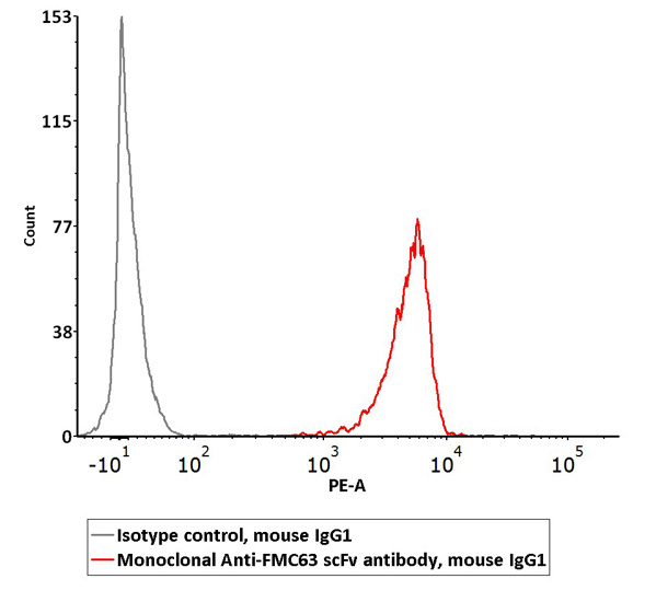
Detection of Anti-CD19 (FMC63) CAR Expression
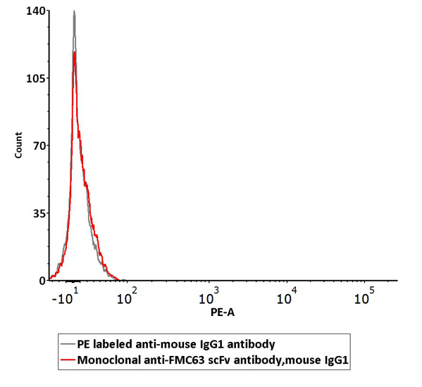
Assessment of Non-specific Binding to 293 Cells
2e5 of FMC63 scFv-based anti-CD19 CAR-293 cells were stained with 100 µL of the working solution of Monoclonal AntiFMC63 scFv Antibody, Mouse IgG1 (Cat. No. FM3-Y45) and isotype control respectively, washed and then followed by PE anti-mouse IgG1 Antibody and analyzed with flow cytometry (QC tested).
Non-specific binding of Monoclonal Anti-FMC63 scFv Antibody (Cat. No. FM3-Y45) to non-transfected 293 cells was determined by flow cytometry. The data showed that Anti-FMC63 scFv Antibody didn’t bind to non-transfected 293 cells.
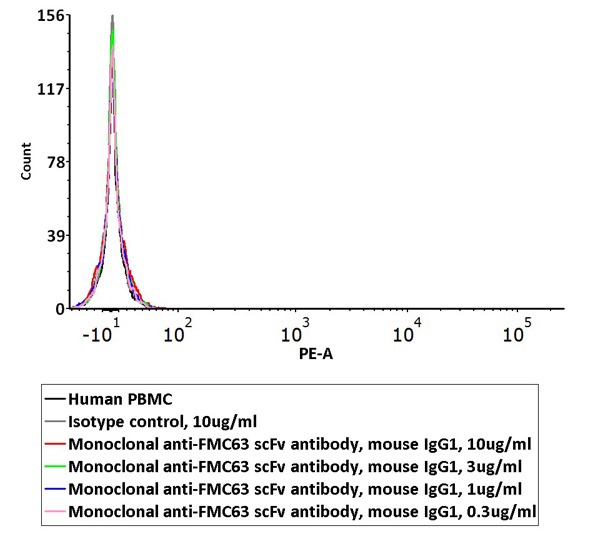
Assessment of Non-specific Binding to PBMCs
Non-specific binding of Monoclonal Anti-FMC63 scFv Antibody (Cat. No. FM3-Y45) to non-transfected human PBMCs was determined by flow cytometry. The data showed that Anti-FMC63 scFv Antibody didn’t bind to non-transfected human PBMCs.
FMC63由来CARの抗原認識領域を高感度、特異的に認識します
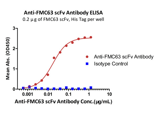
Binding Activity and Specificity Measured by ELISA
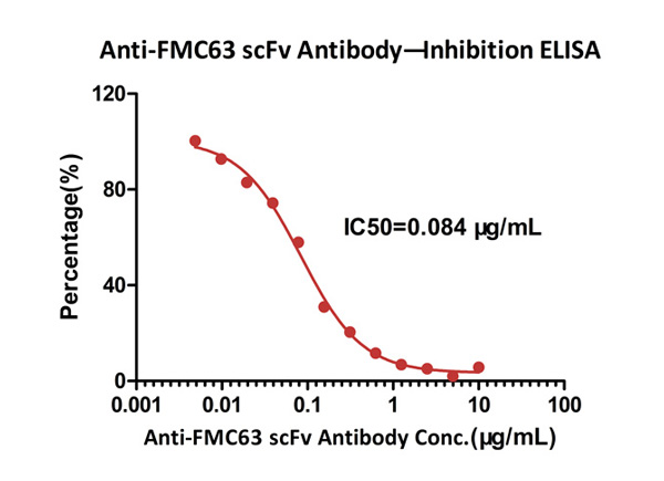
Neutralizing Activity Measured by ELISA
Immobilized FMC63 scFv, His Tag at 2 μg/mL (100 μL/well) can bind Monoclonal Anti-FMC63 scFv Antibody, Mouse IgG1 (Clone Y45) with a linear range of 1-19 ng/mL. Anti-DNP antibody, mouse IgG1 (Cat. No. DNP-M1) was used as an isotype control (QC tested).
ELISA analysis shows that the binding of Human CD19, Fc Tag (Cat. No. CD9-H5251) to FMC63 scFv, His Tag was inhibited by increasing concentration of Monoclonal Anti-FMC63 scFv Antibody, Mouse IgG1 (Clone Y45). The concentration of Human CD19, Fc Tag used is 5 μg/mL (100 μL/well). The IC50 is 0.084 μg/mL (Routinely tested).
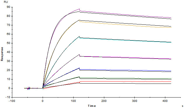
High Affinity Determined by SPR
Monoclonal Anti-FMC63 scFv Antibody, Mouse IgG1 (Cat. No. FM3-Y45) captured on CM5 chip via anti-mouse antibodies surface can bind FMC63 scFv with an affinity constant of 1.08 nM as determined in a SPR assay.
Cynomolgus / Rhesus macaque CD19 (20-292) Protein, His Tag
特長
FACSによる抗CD19 CAR発現の検出
FACSによる抗CD19 CAR発現の検出
1e6 of the anti-CD19 CAR-293 cells were stained with 100 μL of 1:50 dilution (2 μL stock solution in 100 μL FACS buffer) of PE-Labeled Human CD19 (20-291), His Tag (Cat. No. CD9-HP2H3). PE Streptavidin was used as negative control (QC tested).
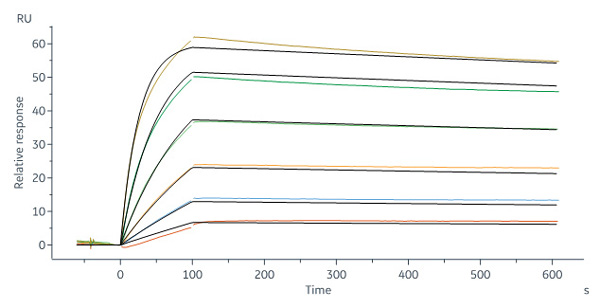
SPRによるアフィニティーの検証
Biotinylated Human CD19 (20-291), His, Avitag (Cat. No. CD9-H82E9) captured on Biotin CAP – Series S sensor Chip can bind FMC63 MAb (mouse lgG2a) with an affinity constant of 0.255 nM as determined in a SPR assay (Biacore 8K) (QC tested).
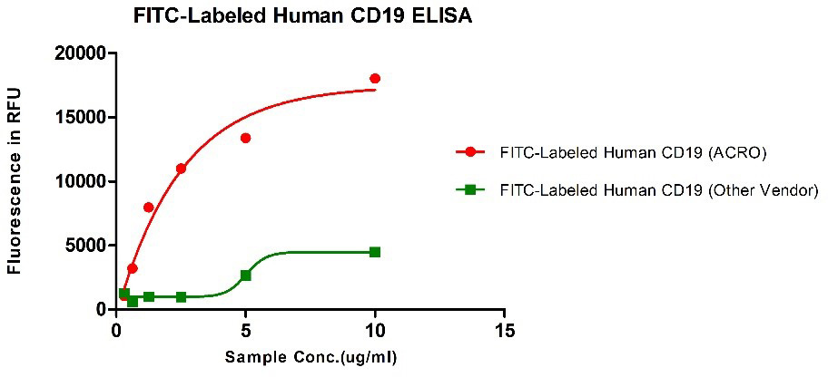
ELISA、FACS、SPRによるアフィニティーの検討
Binding activity of FITC-Labeled Human CD19, His Tag from two different vendors were evaluated in the ELISA analysis against FMC63 Mab. The result showed that ACRO’s FITC-Labeled Human CD19, His Tag has a much higher binding activity than that of the other vendor.
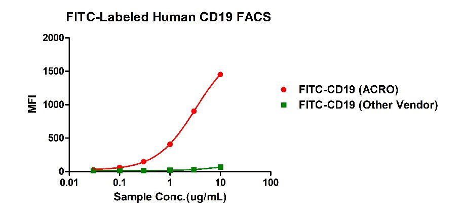
Binding activity of FITC-Labeled Human CD19, His Tag from two different vendors were evaluated in the flow cytometry analysis against anti-CD19-CAR-293 cells. The result showed that ACRO’s FITC-Labeled Human CD19, His Tag has a much higher binding activity than that of the other vendor.
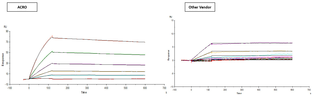
Binding activity of Human CD19, His Tag from two different vendors were evaluated by SPR assay against FMC63 MAb. The result showed that ACRO’s Human CD19, His Tag can bind FMC63 MAb with an affinity constant of 2.95 nM which is much higher than that of the other vendor.
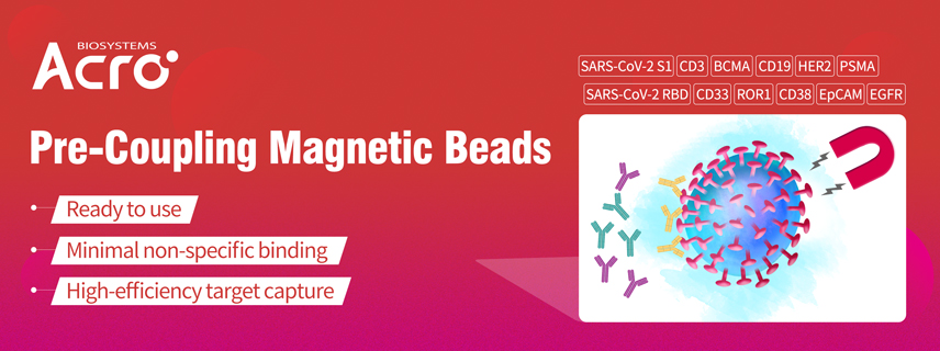
A team from Peking University published their work about the screening of the neutralizing antibodies against SARS-CoV-2 in top journal Cell early this year. In their work, they used RBD and S protein pre-coupled magnetic beads to enrich the RBD binding B cells. Compared to the traditional cell isolation method, the pre-coupling beads help to increase the efficiency to 20-fold.[1]
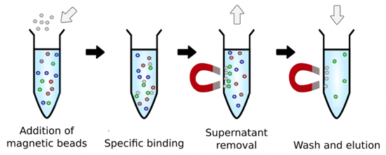
Fig. 1 Workflow of the pre-coupling magnetic beads
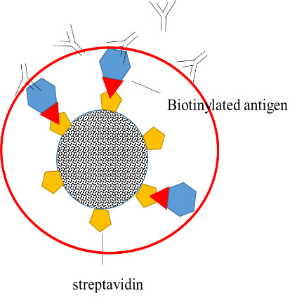
The biotinylated proteins were conjugated to streptavidin magnetic beads. Magnetic particles from alternative suppliers often have random sizes and surface area which could compromise your assay performance. ACRO screened a few different beads and bring this tightly controlled superparamagnetic microspheres to you. The beads are in uniform size, narrow size distribution with large surface area and unique surface coating, which can help you get the best performance and highly reproducible results.
Our pre-coupled beads could capture the molecules which have affinity to the pre-coupled proteins from various assay systems, and make the following testing easy.
>>> Relevant recommendation:
Cat.No. MB-12 Magnetic beads blocking buffer (We recommend using our blocking buffer together with the pre-coupling beads for immunocapture.)
Cat.No. SMB-B01 Magnetic beads™ Streptavidin (We recommend using our non-coupling streptavidin magnetic beads as a control)
Features and Advantages
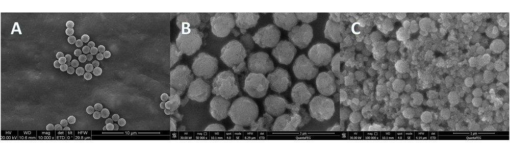
Low non-specific binding
ACRO chose the uniform, better-shaped paramagnetic beads to reduce the non-specific binding.
Fig. 2 TEM data of different magnetic beads. A is ACRO’s magnetic beads; B and C are competitors’ beads.
High efficiency
Our pre-coupling beads are at the level of saturation. A maximum amount of proteins can ensure the best capture performance, which could increase the efficiency of your assay.
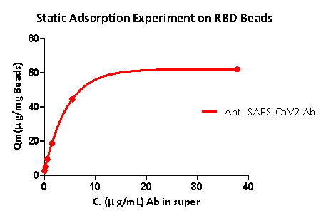
According to the static adsorption experiment result, the RBD pre-coupling magnetic beads can be used to capture the anti-SARS-CoV-2 S1 antibody. RBD pre-coupling amount is greater than 40ug/mg beads.
Fig 3. The binding curve between RBD pre-coupling magnetic beads and anti-SARS-CoV-2 antibody.
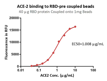
Fig. 4 The binding curve between RBD pre-coupling magnetic beads and ACE2 protein. 0.1 mg of Beads (1mg/mL, 100 L) was washed three times and the supernatant was removed. Antibodies of the corresponding concentration of 100 L (10 g/mL~0.039 g/mL) were added. One hour later,fluorescent labeled secondary antibody was added for another one-hour-reaction, the corresponding Binding signal was detected and the Binding curve as obtained.
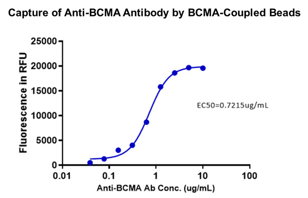
Fig. 5 Use BCMA pre-coupled magnetic beads to capture the anti-BCMA antibody. Anti-BCMA antibody can all be captured before the maximum binding capacity is reached. The PE-labeled secondary antibody is used for detection.
Convenient
Our pre-coupling beads are lyophilized for long term storage. It is ready to use after reconstitution. No need for activation which could greatly save your time and hassle.
Enzyme-linked immunosorbent assays (ELISA) have been around as one of the primary methods of analyte detection for decades. It is regarded as the most accessible and flexible assay formats for the biological assays. ELISA can be used for biopanning, molecule screening, molecule quantification, pharmacokinetic assays and immunogenicity assays, etc. ACRO took the advantages of our own comprehensive list of target protein products, and developed a serial of ready-to-use universal ELISA kits. These ELISA kits will greatly save your time and hassle for your drug development process.
The serum samples contain many factors that may potentially interfere with the indirect ELISA result. This is the major reason for the background issue. We include the diluent buffer in the kit to help solving the problem.
Fig. 2 Determination of serum drug concentration using anti-Her-2 ELISA Kit
High sensitivity and wide detection range
Our ELISA kits utilized an indirect ELISA format. Antigen coating is achieved through the binding between biotin and streptavidin. The sensitivity can reach the level of ng/ml. The detection range is about 2 to 3 logs
Fig.3 Immobilized human LAG-3 protein, Avi Tag (Cat. No. LA3-H82E5) at 1 μg/mL (100 μL/well) can bind Anti-LAG-3 Neutralizing Antibody with a linear range of 4.096-160 ng/mL
Consistent performance
We install rigorous quality control program to ensure the lot-to-lot consistency. Every batch of products are analyzed to meet our internal standards. The product will be released only if all standards are met.
Fig.4 Determination of serum drug concentration using anti-TIGIT ELISA Kit. Consistent results were achieved when perform the assay on various days
Background
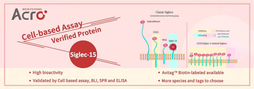
Siglec-15 is a member of the sialic acid-binding Ig-like lectin (Siglecs) family and is expressed in M2 macrophages, myeloid cells, dendritic cells, B cells, and osteoclasts. Siglec-15 can continuously inhibit the activity of T cells and exhibits the main features of normalized cancer immunotherapy. Related studies have shown that Siglec-15 is the major immunosuppressive factor in most PD-L1 negative tumor patients. And the anti-cancer effect of this target is likely to be mutually exclusive with PD-L1. In tumor cells, the expression level of siglec-15 is low when PD-L1 expression is high. For those cancer patients who are not effective in PD-1/PD-L1 treatment, Siglec-15 may bring them a new choice of immunotherapy.
High bioactivity Siglec-15 protein can help drugs development. The bioactivity of Siglec-15 protein developed by ACROBiosystems is verified by Cell based assay/BLI /SPR /ELISA, and the corresponding protocols are provided for free.
Product Features
The bioactivity of Siglec-15 proteins are verified by Cell based assay/ BLI/ SPR/ ELISA, and the protocols are offered.
Cell based assay
BLI
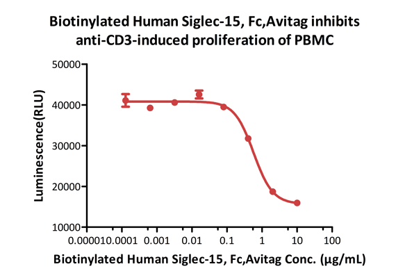
Biotinylated Human Siglec-15, Fc,Avitag (Cat. No. SG5-H82F5) inhibits Anti-CD3-induced proliferation of PBMC. The ED50 for this effect is 0.48-0.57 μg/mL.
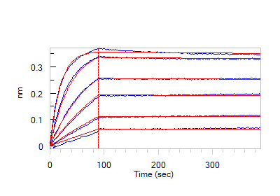
Loaded Anti-Siglec-15 MAb (Mouse IgG) on AMC Biosensor, can bind Human Siglec-15, Fc Tag (Cat. No. SG5-H5253) with an affinity constant of 59.7 pM as determined in BLI assay (ForteBio Octet Red96e).
SPR
ELISA
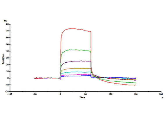
Immobilized Neu5Ac(a2-6)GalNAc-PAA-biotin on SA Chip can bind Human Siglec-15, Fc Tag (Cat. No. SG5-H5253) with an affinity constant of 4.49 μM as determined in a SPR assay (Biacore T200).
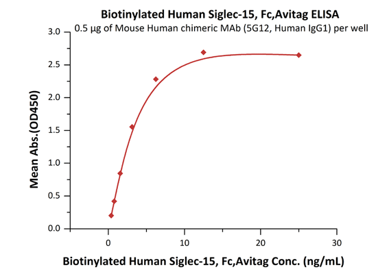
Immobilized Mouse Human chimeric MAb (5G12, Human IgG1) at 5 μg/mL (100 μL/well) can bind Biotinylated Human Siglec-15, Fc,Avitag (Cat. No. SG5-H82F5) with a linear range of 0.4-3 ng/mL.